Modeling development using hydrogels


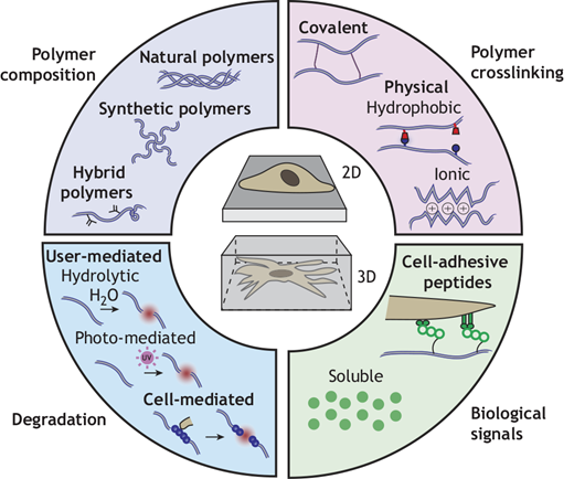
A team of researchers including Robert Mauck, PhD, the Mary Black Ralston Professor of Orthopaedic Surgery and Professor of Bioengineering at the University of Pennsylvania recently covered how engineered hydrogels can serve as in vitro culture platforms to present such signals in a controlled manner and include examples of how they have been used to advance our understanding of developmental biology. You can read more about this research in Development.

Dr. Mauck also co-directs the Program in Musculoskeletal Regeneration at the IRM.  Specific information about current work in the Mauck Lab can be found here.


Reconstitution of human adrenocortical specification and steroidogenesis using induced pluripotent stem cells


Mechanisms leading to adrenal cortex development and steroid synthesis in humans are poorly understood due to the paucity of model systems. In a new study led by Kotaro Sasaki, MD, PhD, in the  School of Veterinary Medicine at the University of Pennsylvania, human fetal adrenal cortex specification processes were recapitulated through stepwise induction of human-induced pluripotent stem cells through posterior intermediate mesoderm-like and adrenocortical progenitor-like states to ultimately generate fetal zone adrenal-cortex-like cells (FZLCs), as evidenced by histomorphological, ultrastructural, and transcriptome features and adrenocorticotropic hormone (ACTH)-independent Δ5 steroid biosynthesis. The researchers believe these findings provide a framework for understanding and reconstituting human adrenocortical development in vitro, paving the way for cell-based therapies of adrenal insufficiency.

Read the full paper from the Sasaki lab in Developmental Cell.

Neurulation of the cynomolgus monkey embryo achieved from 3D blastocyst culture


A joint collaboration between the Plachta Lab, led by Nicolas Plachta, PhD,  the William Richard Gordon President’s Distinguished Professor in Genetics, at the the Perelman School of Medicine at the University of Pennsylvania and the Chinese Academy of Sciences found that understanding human post-implantation development remains a primary challenge due to restrictions on human embryo research, technical limitations of prolonged embryo culture, and inaccessibility to tissue. The study used a 3D prolonged in vitro culture system that supports monkey embryo development up to 25 days postfertilization. This extends primate embryo culture to allow for the study of embryogenesis through advanced gastrulation and early neurulation.

Highlights of the study include:

  • Cynomolgus blastocysts are cultured to d.p.f. 25 within an optimized 3D system
  • Cultured embryos recapitulate developmental features of their in vivo counterparts
  • Early neurulation features are demonstrated in cultured embryos
  • Epigenetic features of three germ layers in primate are unveiled in cultured embryos

Read the full paper, as published in Cell, here.

Human embryo live imaging reveals nuclear DNA shedding during blastocyst expansion and biopsy

A team of researchers including Nicolas Plachta, PhD, from the Perelman School of Medicine at the University of Pennsylvania identified that proper preimplantation development is essential to assemble a blastocyst capable of implantation. Live imaging has uncovered major events driving early development in mouse embryos; yet, studies in humans have been limited by restrictions on genetic manipulation and lack of imaging approaches. Researchers overcame this barrier by combining fluorescent dyes with live imaging to reveal the dynamics of chromosome segregation, compaction, polarization, blastocyst formation, and hatching in the human embryo.

The work reveals distinct processes underlying human development compared with mouse and suggests that aneuploidies in human embryos may not only originate from chromosome segregation errors during mitosis but also from nuclear DNA shedding.

Read the full paper, as published in Cell, here.

1 2 3 6