Distinct first responder cells lead the skin’s wound healing response

Under normal conditions, a menagerie of separate cell populations work to maintain skin health. However, injury prompts these isolated pools of cells to exit their individual niches and re-epithelialize the epidermis (the surface skin layer). This raises important questions about which cells react first and how the body primes them for rapid injury response.

A study by the Rompolas lab published this week in Cell Stem Cell provides a closer look at how mice maintain these pools of cells and how the cells respond after injury. Led by postdoctoral researcher Sixia Huang, the team used live cell imaging to confirm that that cells expressing Lgr6 serve as first responders that proliferate and initiate epithelial repair following injury. Moreover, the researchers show that targeted destruction of Lgr6+ cells slows the injury response by requiring other stem cells to respond.

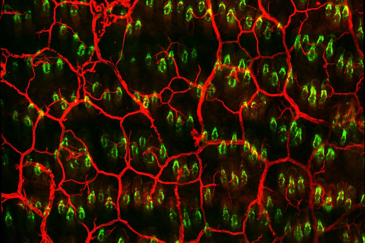
So what makes Lgr6+ cells step up first? Consistent with previous data showing that Lgr6 expression by keratinocytes depends on skin innervation, the researchers find that sensory nerves contact the pool of Lgr6+ stem cells and are required for normal cellular dynamics post-injury. This nerve interaction regulates Lgr6+ cell identity and fate by regulating the expression of other genes within the cells, something that is drastically altered if nerves are lost.

Altogether, these results shed light on how the nervous system crosstalks with stem cells in the body. Rather than simply serve as pain receptors, cutaneous nerves prime Lgr6+ cells to function as first responders to injury. These observations may shed light on why patients suffering medical conditions associated with neuropathies, like diabetes, may also suffer from diminished wound healing.

Photo courtesy of Sixia Huang

The immune link between a leaky blood-brain barrier and schizophrenia

Like a stern bodyguard for the central nervous system, the blood-brain barrier keeps out anything that could lead to disease and dangerous inflammation—at least when all is functioning normally.

That may not be the case in people with schizophrenia and other mental disorders, suggest new findings from a team led by researchers from the School of Veterinary Medicine, Perelman School of Medicine, and Children’s Hospital of Philadelphia (CHOP). In these individuals, a more permissive barrier appears to allow the immune system to get improperly involved in the central nervous system, the researchers showed. The inflammation that arises likely contributes to the clinical manifestations of neuropsychiatric conditions.

Read more about how Jorge Alvarez, Stewart Anderson and their team used patient-derived stem cells and mouse models to examine the role of immune cells in schizophrenia in Penn Today.

Arcuate organoids to study development and disease of the hypothalamus

Human brain organoids are remarkable platforms for modeling features of human brain development and diseases. Building on methods to generate organoids to model different brain regions such as the cortex and the midbrain, researchers at the Perelman School of Medicine at the University of Pennsylvania have generated the first organoids of the arcuate nucleus (ARC), an essential structure in the hypothalamus that sends signals of hunger and feeling full. This part of the hypothalamus exhibits a tremendous amount of cell diversity, and is far more complex than previously modeled parts of the brain.

In a recent paper in Cell Stem Cell, researchers at Penn report generating arcuate organoids (ARCOs) that model the ARC of the hypothalamus. Previous studies have generated 2D hypothalamic-like neurons and 3D hypothalamic organoids from human induced pluripotent stem cells (iPSCs). However, no protocols previously existed to generate hypothalamus nucleus-specific organoids.

Read more about this work from Guo-li Ming‘s lab in Penn Medicine News.

New research uncovers distinct roles for corneal stem cells

How does the cornea maintain its rapid pace of cell turnover—and how does the body regenerate it after injury? A research team led by Pantelis Rompolas, Assistant Professor of Dermatology and Olivia Farrelly, a Ph.D. candidate in the Cell and Molecular Biology Graduate Group, uncovered exciting new clues by identifying separate stem cell pools for maintenance and injury repair.

The investigators used two-photon live imaging in mice to explore the cell dynamics of the corneal limbus, an important adult stem cell niche that maintains the cornea. They find that stem cells display different roles and fates depending on location: cells in the inner limbus primarily maintain the cornea during normal conditions while cells of the outer limbus step in to help regenerate the cornea after injury.

These findings reconcile prior observations in the field regarding how the limbus contributes to eye health and inform ongoing attempts to develop cell therapies for ocular diseases. You can read more about this research in Cell Stem Cell.