Pathway behind muscle breakdowns in Duchenne muscular dystrophy discovered

An overactive genetic pathway in muscle stem cells was found to shorten the ends of chromosomes, called telomeres, resulting in DNA damage that impedes the normal healing response, according to a new study by researchers in the Perelman School of Medicine at the University of Pennsylvania. The researchers believe this finding unveils the body’s origin point for the chronic muscle injuries associated with diseases like Duchenne muscular dystrophy. This work was published today in Cell Reports.

The specific pathway the researchers studied, NF-kB, is tied both to DNA transcription and inflammation response, among other things. It is the first genetic pathway found to directly affect telomere shortening. Shortened telomeres were previously identified as a key feature of patients with Duchenne muscular dystrophy, a severe muscle disease characterized by extensive muscle injury and repair.

Read more about this work from the Mourkioti lab in Penn Medicine News.

The origin of reproductive organs

Early in human development, during the first trimester of gestation, a fetus may have XX or XY chromosomes that indicate its sex. Yet at this stage a mass of cells known as the bipotential gonad that ultimately develops into either ovaries or testes has yet to commit to its final destiny.

While researchers had studied the steps that go into the later stages of this process, little has been known about the precursors of the bipotential gonad. In a new study published in Cell Reports and co-led by Kotaro Sasaki of Penn’s School of Veterinary Medicine, an international team lays out the detailed development of this key facet of sexual determination in two mammalian models.

Read more in Penn Today

Photo credit: Kotaro Sasaki

Research Recap: Scar skipping, filament foretelling, fracture fixing, and more

Recent preprints and publications from IRM researchers. This month: a path toward scar-less healing, physical drivers of cell fate, factors that influence fracture repair, and a method to follow a cell’s RNA through time. (Image from Plachta lab)

Using an old cream to prevent new scars

Mammals tend to repair wounds by forming scars. While helpful for preventing further injury or infection, fibrotic scars don’t have all the cell types normally found in skin, resulting in changed appearance and function. Researchers in the Leung and Cotsarelis laboratories have demonstrated that topical imiquimod, a cream used to treat skin cancer and warts, can prompt mice to skip scarring and initiate regenerative healing instead. Their results suggest that imiquimod activates TRPA1, a nociceptor (“pain receptor”) that in turn induces an immune response mediated by γδ T cells essential for scar-less healing. These results set the stage for clinical trials in humans. (Science Immunology; read more in Forbes).

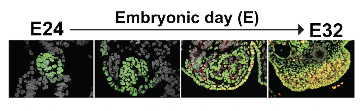
Keratin helps embryos sort inside from outside

Every cell in the body ultimately comes from the embryo. But how mammalian embyonic cells choose the right “fates,” ensuring that the right cell types wind up in the right places, is still an open question. A new paper from the Plachta lab provides evidence that keratins, a family of proteins that provide cells with structure, act as markers of an important cell fate decision. The team used live cell imaging to show that keratin appears in only certain cells at the 8-cell stage. These keratin filaments are then inherited by a subset of daughter cells after cell division, imbuing these cells with structural “memory” that positions them on the outer edge of the embryo. As development progresses, these outer cells become the trophectoderm, a layer of nourishing tissue that later forms the placenta. (Nature; read more in News and Views).

Setting the stage for proper bone healing

As anyone who has suffered a major accident can tell you, bone healing is a remarkable process. In response to fractures, progenitor cells in the lining (periosteum) of long bones kick into gear, expanding in number and differentiating into cartilage- and bone-producing cells. A new paper from the Boerckel and Qin labs demonstrates that two transcription factors, YAP and TAZ, are required for proper osteoblast (bone-forming cell) development in response to injury in mice. By selectively controlling YAP and TAZ expression during mouse development and adulthood, the researchers showed that these proteins are required for both the expansion of periosteum progenitor cells and their differentiation into osteoblasts after fracture. In contrast, YAP and TAZ loss in adult mice had little effect on cartilage formation. Future work will help researchers develop therapies for fractures that don’t heal properly. (Journal of Bone and Mineral Research)

Sorting new RNA from old at the single cell level

Information about a cell’s mRNA is very insightful. With it, we can see which genes are turned on or off and link these patterns to unique cell types and states. Unfortunately, standard methods for gathering these data provide only snapshots in time, preventing closer analysis of ever-changing mRNA levels. To overcome this limitation, the Wu lab and collaborators adapted several labeling, sorting, and chemical techniques into “single-cell metabolically labeled new RNA tagging sequencing,” or scNT-seq. After showing that scNT-seq can distinguish newly transcribed from pre-existing mRNA, the team turned scNT-seq loose to see how stem cells regulate mRNA during transitions between states, including rare totipotent two-cell embryo (2C)-like stem cell states. The researchers hope scNT-seq will help others understand highly dynamic biological systems. (Nature Methods)


Research Recap: Following the footsteps of metastatic cancer, blood development, and COVID-19

Recent preprints and publications from IRM researchers. This month: a new technique for tracing cancer cells back to the source, another step toward producing patient blood stem cells outside of the body, and a search for coronavirus targets in the brain that leverages insights from other viral fights.

Which cancer cells go rogue?

Why do some cancerous cells metastasize to other parts of the body, often with grave consequences for health? In a preprint posted this month, researchers from the Lengner and Stanger labs use a new technique, macsGESTALT, to study the origin of metastatic pancreatic cancer cells. After stimulating tumor growth in mice with an injection of specially engineered pancreatic cells, the researchers collected information on tens-of-thousands of cells away from the injection site. Using sophisticated computational analysis, they were able to trace the “lineage” of these colonizing cells back to specific cells in the original tumor. So what makes a cell more likely to take up root somewhere else? According to the researchers, led by MD-PhD student Kamen Simeonov, metastatic cells overwhelmingly originate from a subgroup of highly aggressive tumor cells that progress mostly—but not fully—through a pathway known as epithelial–mesenchymal transition (EMT). They anticipate that the macsGESTALT technique will help other teams answer questions about cancer biology and the development of stem cells into functional tissues. (bioRxiv)

Finding a blood-making bottleneck

Hematopoietic stem cells (HSCs) give rise to the blood cells that carry oxygen throughout the body and fight infections. For patients of many hematological diseases, an HSC transplant restarts the blood production system after it is destroyed by chemotherapy. But there is a catch: successful transplants require matching donors, limiting the number of patients able to receive this therapy. To get around this limitation, scientists are developing methods to grow large numbers of compatible HSCs outside of the body. As a step toward this goal, researchers from Kai Tan and Nancy Speck’s laboratories profiled nearly 40,000 rare single cells from sites of HSC formation in embryonic mouse arteries over a three day window. Using a pair of methods to watch which genes get “turned on,” or expressed, during this crucial period, the researchers found a bottleneck along the pathway by which cells transition into HSCs. Cells exit this bottleneck—termed the “pre-hemogenic endothelial”, or “pre-HE,” stage—when RUNX1, a gene known to be critical for HSC development, is expressed. By pinpointing when RUNX1 becomes vital and characterizing different cell populations later in the three-day period, the team uncovered important conditions for growing HSCs in the laboratory. (Blood)

Probing for coronavirus weak spots in the brain

Although we know COVD-19 as a respiratory illness, its effects go well beyond the lungs. Patients can suffer from a variety of symptoms—including neurological issues such as dizziness and confusion—that suggest a propensity for SARS-CoV-2 to infect cells throughout the body. To search for potential viral targets in the brain, researchers from the Ming and Song labs took advantage of a system previously used to understand the behavior of Zika virus: organoids cultured from human-induced pluripotent stem cells (hiPSCs). Organoids use a combination of hiPSC-derived cell types to mimic the three-dimensional structure of actual human organs. After growing organoid models of the cerebral cortex, hippocampus, hypothalamus, and midbrain, the researchers exposed each “minibrains” to SARS-CoV-2. Their results suggest that choroid plexus epithelial cells, which line the blood/cerebral spinal fluid (CSF) barrier, are prone to high levels of infection, offering clues for further exploration of COVID-19’s impact on the brain. (bioRxiv, final manuscript in Cell Stem Cell)